
The advanced neurological testing procedures listed below allow us to pinpoint brain impairments that may be responsible for your migraines
Video Nystagmography VNG
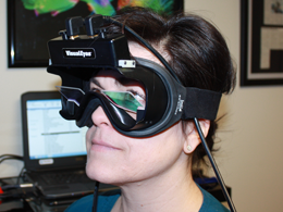
VNG is a computerized system used in the assessment of the oculomotor system (nervous system control of eye movement) and the function of those areas of your brain controlling these movements.
The VNG test is comfortably performed in our office. We simply place special goggles with an infrared camera on your head to record and measure your eye movements in both light and dark conditions.
Specific areas and pathways of the brain are activated during different types of eye movements. Many of the eye movement areas of the brain are located in the brain stem close to the area of dysfunction in migraines.
During the test, computer software is recording and graphing your precise eye movements as you follow the visual target with your eyes. By measuring your recorded eye movements and displaying them on the computer screen, you can see your own eye movements and how they compare to normal measurements.
The following eye movements are measured by VNG:
- Gaze Stability: It is essential that the brain stabilizes the gaze of the eyes for proper equilibrium and balance. One of the most common ocular motor deficiencies seen in neurological disorders is poor gaze stability. This can be seen on the VNG tracing.
- Pursuits: Pursuits are slow and steady eye movements that allow you to track a moving object. Good (smooth) pursuits are essential to everyday functions. The pursuit mechanism is commonly altered in patients that have neurological conditions.
- Saccades: Saccades are fast eye movements used to shift a person’s gaze from one object to another. Saccades need to be fast and accurate. Sub-optimal brain function can alter the speed and accuracy of a person’s saccades.
- Optokinetics (OPK): This is an essential eye movement that allows for visual function as the surrounding visual environment passes by. When these are dysfunctional it can indicate poor brain function.
The VNG eye movement tracings provide invaluable neurologic information concerning specific areas of your brain affected. We are experts at analysing these specific eye movements and determining if they have been disrupted in their proper function. We correlate the VNG findings with our other testing and examination procedures to develop a specific therapeutic program for each individual.
Saccadometry
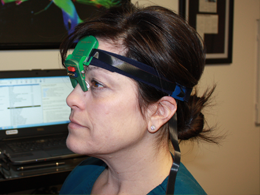
We measure your saccades using saccadometry. Saccades are quick movements of your eyes that allow you to voluntarily move your eyes quickly from one object to another or to react by moving your eyes to a suddenly appearing target reflexively. Saccades can be measured using a device called a saccadometer. The saccadometer is strapped around a patient’s head using an elastic headband. Within the unit, there is a laser that projects targets onto the wall that a patient will look at quickly or saccade to. The saccadometer will measure the eye movements at the same time.
Studies have shown this test to be a very accurate way to detect brain dysfunction caused by injury or neurological disorders. This test cannot be altered voluntarily by the patient because you cannot consciously control the speed of your eye movement during each saccade; your eyes move as fast as they can. The purpose of your saccadic eye movement is to aim the part of your eye with the best vision (fovea) at the thing you want to see most, and then hold your eye there as you examine it.
Saccades are the only human physiological function that uses every area of the brain. It is for this reason that we can evaluate a person’s saccades to determine what areas of the brain are not functioning up to par.
Some of the brain areas you use in controlling saccades involves three main cortical areas: the frontal eye fields, the parietal eye fields, and the supplementary eye fields; and several subcortical areas in the basal ganglia, thalamus and brainstem.
Saccadometry very precisely measures these aspects of saccades:
- Latency: This is the reaction time taken for the saccade to fire or activate after a new stimulus or target is presented. A normal reaction time should be less than 200ms. People who have had a concussion or suffer from neurodegenerative disorders such as Alzheimer’s and Parkinson’s disease have pathologically long saccadic latency times that are greater than 200ms.
- Velocity: The speed of a saccade is highly related to health of areas in the brain stem commonly affected in neurological disorders.
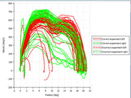 Accuracy: The ability to make the eyes stop and land directly on the target they are saccading to or looking toward is essential to a person functioning in their environment accurately. If the saccade overshoots or undershoots the target it may indicate that the cerebellum or the area of the brain that makes the environmental grid may have been affected by injury or neurological disorders. The saccadometer computerized graph shows leftward saccades that are generated by the right brain hemisphere as red and the rightward saccades that are generated by the left brain as green. This can be very useful in determining what side of the brain is most affected.